Diagnostic Neuroimaging
Multimodality imaging techniques for diagnosis of blood vessel disorders of the brain and spine, including cerebral arteriography, venography, and myelography.
You can jump directly to the section of interest by clicking on the links below.
Angiography
- Carotid/Cerebral Angiography
- Spinal Arteriography
- Venography
- Myelography
- Wada Testing
- Inferior Petrosal Sinus Sampling
Carotid/Cerebral Angiography
 Cerebral arteriography is a form of angiography which shows images of blood vessels in and around the brain, thereby allowing detection of abnormalities such as aneurysms, arteriovenous malformations, fistulas, and tumors.
Typically a catheter is inserted into a large artery (such as the femoral, brachial, or radial artery) and threaded through the circulatory system to the carotid, vertebral, or subclavian artery, where a contrast agent (dye) is injected. A series of radiographs are taken as the contrast agent spreads through the brain’s arterial and venous systems.
For some applications, and in particular when dymanic information is useful or necessary, this method yields better diagnostic information than less invasive methods, such as computed tomography angiography (CTA) and magnetic resonance angiography (MRA).
In addition, cerebral angiography allows certain treatments to be performed immediately, based on its findings. If, for example, the images reveal an aneurysm, metal coils may be introduced through the catheter already in place and maneuvered to the site of aneurysm; over time these coils encourage formation of connective tissue at the site, strengthening the vessel walls.
Spinal Arteriography
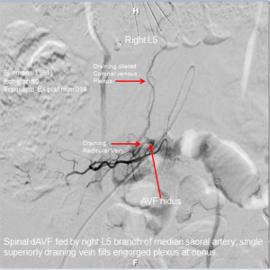
 Spinal arteriography is a form of angiography which shows images of blood vessels in and around the spine, thereby allowing detection of abnormalities such as spinal dural fistulas, spinal arteriovenous malformations, and tumors.
Venography
Myelography
 Myelography is a type of radiographic examination that uses a contrast medium to detect pathology of the spinal cord, including the location of spinal narrowing, cysts, and tumors. The procedure involves insertion of needle into the spinal canal followed by injection of contrast (dye) and then several X-ray images in different projections. A myelogram may help to find the cause of pain not found by an MRI or CT.
Wada Testing
 The Wada test, also known as the “intracarotidsodiumamobarbital procedure” (ISAP), is used to establish cerebral language and memory representation of each hemisphere. Abarbiturate (which is usually sodium amobarbital) is introduced into one of the internal carotid arteries via an intra-arterial catheter. The drug is injected into one hemisphere at a time. The effect is to shut down any language and/or memory function in that hemisphere in order to evaluate the other hemisphere (“half of the brain”). Then a neuropsychologist will perform a series of memory and language related tests on that side of the brain. After a few minutes the medicine wears off, and that side of the brain wakes up. The procedure is then performed on the other side, and the neuropsychologic tests are repeated. An epileptologist interprets the test results.
Inferior Petrosal Sinus Sampling
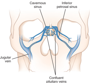
 Inferior petrosal sinus sampling (IPSS) is a rigorous approach to the diagnosis of Cushing’s disease. It tests to see the source of the raised ACTH levels in a patient with Cushingoid features. To technically describe this minimally invasive procedure, a catheter is placed in the inferior petrosal sinus, and multiple blood samples are taken. Hormones (ie. Corticotropin or Vasopressin analogues) are injected, and samples are taken to measure the pituitary’s response to the injected hormones. This tests the pituitary function. In short, the inferior petrosal sinus (IPS) is where the pituitary gland drains. Therefore, a sample from the IPS showing raised ACTH compared to the periphery suggests that the cause of Cushing’s is the pituitary gland. By definition, this is Cushing’s disease and may be treatable by pituitary surgery. The test is performed following a low dose dexamethasone suppression test. Increasingly, it is known as the gold-standard method for diagnosing Cushing’s disease.